

Foot Orthotics and Osteoarthritis: Their Relationship
What is osteoarthritis?
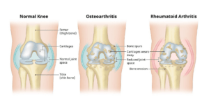
Knee osteoarthritis is a condition characterized with degradation of bone and cartilage within the
joint leading to an abnormal inflammatory response. The inflammation, as well as friction and abnormal loading, results in bone growths calls ‘osteophytes’. These growths combined with reduced joint space, change the normal movement of the joint and results in a high amount of pain.
The image below explains the difference between a healthy looking joint and arthritic joint:
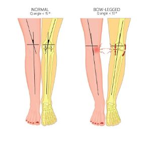
When these changes occur, the joint undergoes a different amount of force, which can cause deformities such as bow-leg deformity or Varus knee. This biomechanical change occurs in mid to late stages of the disease and is worse in patients who already have a varus alignment in their knees.
As a result of these deformities, the medial compartment receives a higher amount of force which means it has a higher amount of compression.
What does this have to do with your feet?
 These changes do not just stay confined to the knees. In many instances, the foot is affected too. The subtalar joint in the foot rolls inwards, resulting in overpronation or ‘flat foot’. As the foot
rolls in, there is more contact with the ground. More area of contact of foot to ground = more force = more force from ground upwards as we know from Newton’s first third law of motion: for every action, there is an equal and opposite reaction.
The force that comes up from the ground is called ground reaction force (GRF). A small amount of medial rotation happens between the thigh and the leg bone when walking, which is known as the knee adduction moment (KAM). According to research, people with moderate to severe knee OA have higher KAM.
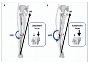 In the image to the right, Figure A represents a normal joint, Figure B represents a OA knee. When there is a varus deformity in the knee along with overpronated foot, the KAM and GRF which causes more compression of the medial joint space.
Treatment:
All this can be reversed by 30-50% by using foot orthotics or a wedged insole. Foot orthotics reduce the GRF therefore reduces the KAM and thus the compressive loads on the knee.
For a long time, foot posture has been considered to contribute to the development of a range of lower limb conditions as it may change the mechanical alignment and dynamic function of the lower limb. Therefore, special attention has been given to foot orthoses and modifications as a non-operative treatment of knee OA.
People with medial compartment knee OA exhibit a more pronated foot type. It is recommended that the assessment of patients with knee OA should include simple foot posture measures. Clinicians should consider the potential influence of foot structure and function on the efficacy of foot orthoses in the management of medial compartment knee OA.
As well as selling premade orthoses, we do custom made orthotics here at PhysiCo. All podiatry done by Eryny Saman, for more info head to her page on our website: https://www.physicocityphysio.com.au/team/#1687820283412-4b8c7616-5d38
References:
Levinger P, Menz HB, Fotoohabadi MR, Feller JA, Bartlett JR, Bergman NR. Foot posture in people
with medial compartment knee osteoarthritis. J Foot Ankle Res. 2010 Dec 16;3:29. doi:
10.1186/1757-1146-3-29. PMID: 21162748; PMCID: PMC3020154. https://pubmed.ncbi.nlm.nih.gov/21162748/
Hall, M., Bennell, K. L., Wrigley, T. V., Metcalf, B. R., Campbell, P. K., Kasza, J., Paterson, K. L., Hunter,
D. J., & Hinman, R. S. (2017). The knee adduction moment and knee osteoarthritis symptoms:
relationships according to radiographic disease severity. Osteoarthritis and cartilage, 25(1), 34–41.
https://doi.org/10.1016/j.joca.2016.08.014 https://pubmed.ncbi.nlm.nih.gov/27616685/